
Women's health
The health of the woman is at the origin of our history.
En Centro de Diagnóstico Granada contamos con los equipos más innovadores para la detección de Cáncer de mama, y de otras patologías de la mujer, como mamografía digital 3D con tomosintesis, ecografía de alta definición, Densitometría ósea, Biopsia Estereotáxica y resonancia magnética de mamas.
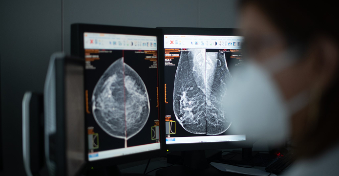
EARLY DETECTION OF BREAST CANCER
Our company was born as Centre for Early Diagnosis of Breast Cancerwith an extensive program of collecting epidemiological data, and of the risks of personal, family and environmental, that are still in force.
Since then it has been especially careful of this pathology, performing and diagnosing more than 80,000 annual mammograms about in our unit of the breast, with a complete process of study:
● Realization of the exploration monographic.
● Realization of ultrasound mammary complementary in the necessary cases.
● Report of radiologic findings in all cases.
● Jacks sample by means of Biopsy in Table Prone Digital.
● Punctures eco tours.
● Placement of harpoons and markers breast.
● Studies Magnetic resonance imaging of Breasts with processing workstation and localization of lesions for biopsy in RM.
● Evaluation and assessment of new technologies.
Esta función preventiva del cáncer de mama forma parte de nuestro ADN y con más de 35 años de experiencia y con medios de última tecnología la llevamos a cabo de la forma más eficaz. Disponemos de un equipo propio de Médicos Especialistas en Radiodiagnóstico expertos y con una formación continuada y de
Técnicos Especialistas en Radiodiagnóstico con una enorme preparación y entrenamiento.
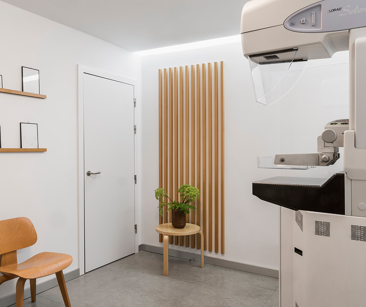
MAMMOGRAPHY AND BREAST ULTRASOUND
Llevamos muchos años trabajando de forma incesante en la detección precoz del cáncer de mama, implementando y aplicando potentes herramientas para el diagnóstico, manteniendo la misma filosofía con la que partimos en nuestros inicios.
Disponemos de sistemas de última generación con adaptación automática a la mama que nos permite un ajuste automático de dosis, de este modo conseguimos evitar repeticiones y reducir hasta un 60% la dosis de radiación empleada en esta prueba.
3D ULTRASOUND AND 4D
The Centro de Diagnóstico Granada features scanners high tech with up to seven probes, three of them in 4D, which allow to obtain three-dimensional images in real-time. Studies are conducted abdomen, pelvis, thyroid, prostate, extremities, skull and hips childin addition obstetric ultrasound 3D and 4D, a technique in which the center was a pioneer in Granada.
The ultrasound three-dimensional offers the possibility of observing the organs from various planes, which improves the detection of pathologies and their relationship with nearby structures. In obstetrics, in addition to the emotional value of seeing the face of the baby, this technique provides detailed information about his internal development and external. If it requires a more in-depth assessment may be supplemented with other tests, such as magnetic resonance imaging.
BONE DENSITOMETRY
The bone densitometry it is a test that measures the mineral density of the bones to detect osteopenia or osteoporosis, pathologies that increase the risk of fractures, especially in women after the menopause. Is done through a system of Densitometry Dual Energy (DEXA), which uses X-rays of high and low energy to differentiate the bone tissue of the soft and accurately calculate the density.
El resultado se compara con los valores normales según edad, sexo y raza, representándose en una gráfica que permite clasificar los resultados como normales, osteopenia u osteoporosis, según los criterios de la OMS. El informe también incluye el porcentaje de masa ósea y una estimación del riesgo de fractura en columna y cadera (sin riesgo, moderado o alto).
Is recommended annually after the menopause or before in people with risk factors (prolonged inactivity, eating disorders, diseases that affect the bone). It is a safe exploration, with a minimal exposure to radiation, even lower than that of a conventional x-ray.
BIOPSY STEREOTACTIC
Before the suspicion or diagnosis of breast cancer, no palpable, it is necessary to perform a biopsy to obtain a tissue sample from you to confirm the nature of the injury. In the Centro de Diagnóstico Granada it uses the most advanced technology and secure: the vacuum-assisted biopsy (BAV) in table prone. In this procedure, the patient lies face down with the breast suspended through a hole, which allows the medical team to access, with accuracy to the area of interest.
The biopsy is performed with local anesthesia, so ambulatorywithout the need of stitches or hospitalization, and leaves minimal discomfort or scarring. The needle of the vacuum allows you to get several samples with a single insertion, avoiding repetition of punctures and ensuring a complete diagnosis and reliable.
Esta técnica ha sustituido en gran medida a la biopsia quirúrgica, ya que ofrece una mayor precisión, menor riesgo y una recuperación más rápida, siendo la opción preferida en países avanzados. Está indicada cuando en la mamografía aparecen microcalcificaciones o masas sospechosas no palpables, o cuando se prefiere un método no quirúrgico de evaluación.
El procedimiento dura entre 30 y 60 minutos, y las muestras obtenidas son analizadas por un patólogo especializado, que emite el resultado en pocos días. Entre sus ventajas destacan la rapidez, fiabilidad diagnóstica, menor coste y pronta reincorporación a la actividad normal, con un riesgo muy bajo de complicaciones como hematomas o infecciones. En conjunto, la BAV es una técnica eficaz, segura y mínimamente invasiva para el diagnóstico precoz del cáncer de mama.
MAGNETIC RESONANCE IMAGING OF THE BREAST
The magnetic resonance imaging of the breast it has been a big step forward in diagnostic imaging. Uses magnetic fields to generate internal signals of the body, captured by antennas special transform that information into images with high precision. In this technique, the breasts are kept in suspension and without compression, which improves the comfort and quality of the study.
Customarily used intravenous contrastbecause the malignant cells capture it with greater intensity, allowing you to identify areas suspicious. The images are processed in workstations advanced, which provide reconstructions three-dimensional and graphics capture detailed, which requires a thorough analysis by the specialist.
The major associations in oncology recommend this exploration in cases of carcinoma in situ, lobular hyperplasia, atypical, mammograms dense, or a personal history of breast cancer. It is also advisable for women with genetic mutations BRCA or inherited syndromes of high-risk (Li-Fraumeni, Cowden, Bannayan-Riley-Ruvalcaba), as well as in patients with high riskaccording to the assessment tools available medical.
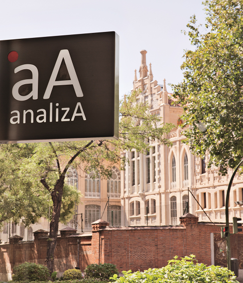
Brands
La incorporación del Centro de Diagnóstico Granada a Analiza amplía nuestras capacidades en diagnóstico por imagen. Su reconocida trayectoria y tecnología avanzada se integran en nuestra red para ofrecer una visión más completa de la salud, alineada con nuestro modelo de diagnóstico integral.

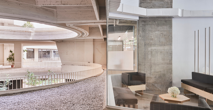
Centro de Diagnóstico Granada
Specialists in clinical analysis and diagnosis by imaging with personalized attention.
