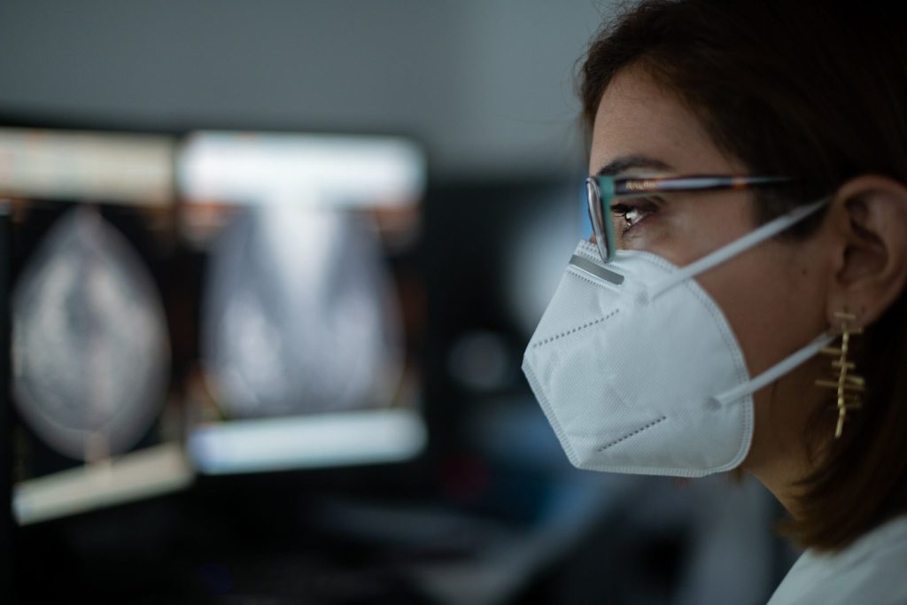

EARLY DETECTION OF BREAST CANCER
Our company was born as Centre for Early Diagnosis of Breast Cancerwith an extensive program of collecting epidemiological data, and of the risks of personal, family and environmental, that are still in force.
Since then it has been especially careful of this pathology, performing and diagnosing more than 80,000 annual mammograms about in our unit of the breast, with a complete process of study:
● Realization of the exploration monographic.
● Realization of ultrasound mammary complementary in the necessary cases.
● Report of radiologic findings in all cases.
● Jacks sample by means of Biopsy in Table Prone Digital.
● Punctures eco tours.
● Placement of harpoons and markers breast.
● Studies Magnetic resonance imaging of Breasts with processing workstation and localization of lesions for biopsy in RM.
● Evaluation and assessment of new technologies.
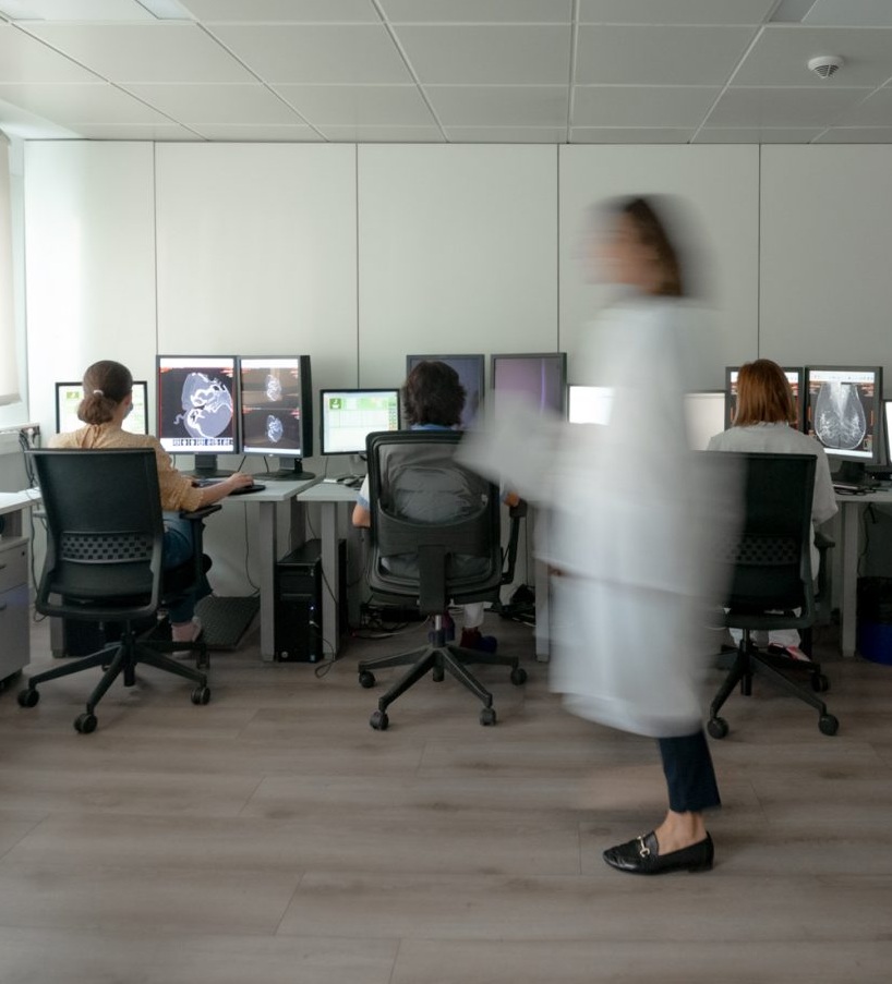
This preventive function of breast cancer is part of our DNA and more than 35 years of experience and with means of the latest technology we bring it out in the most efficient manner. We have a its own team of Medical Specialists in Radiology experts and with continuous training and Technical Specialists in diagnostic Radiology with a huge preparation and training.
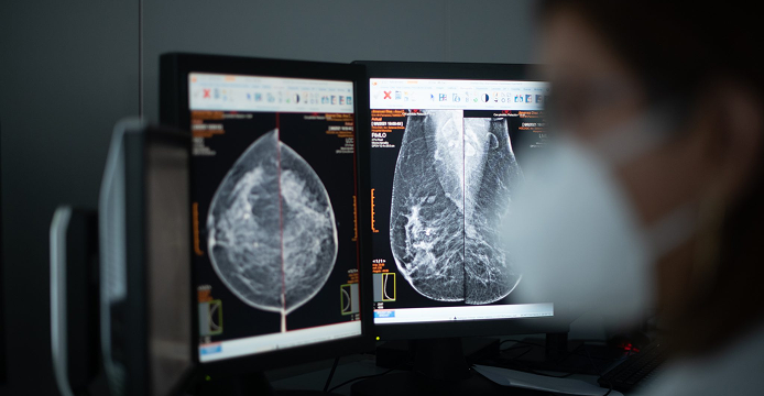
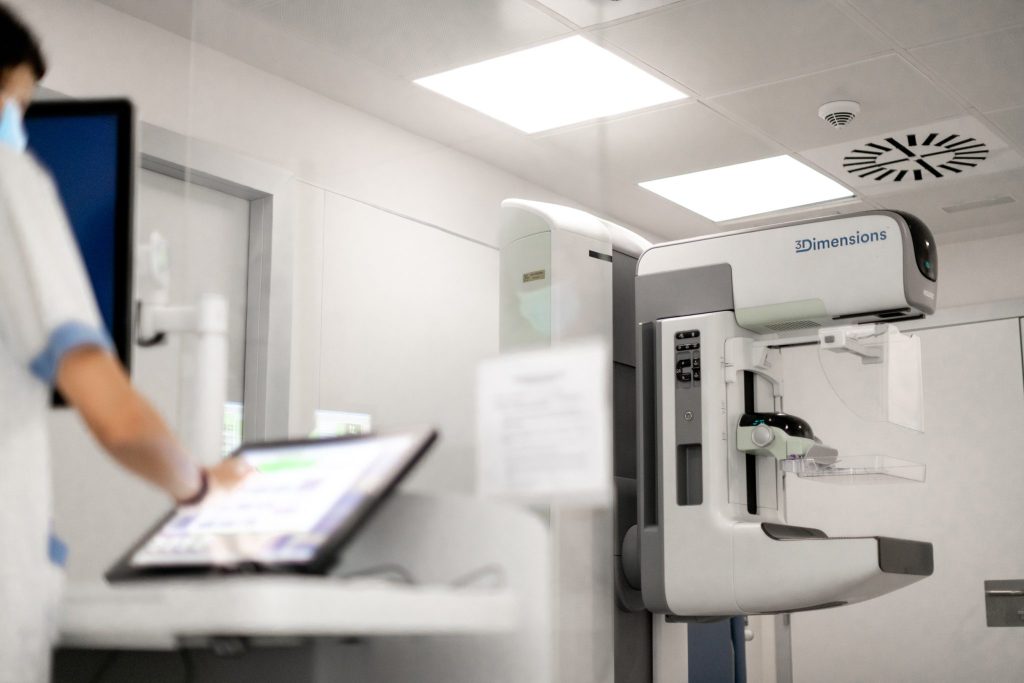
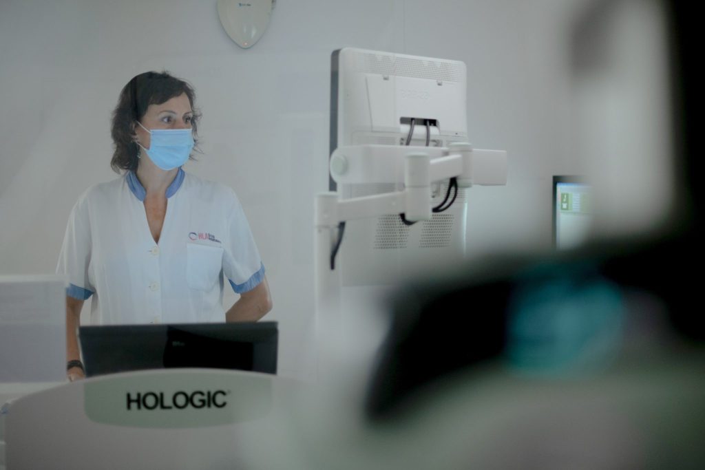
MAMMOGRAPHY AND BREAST ULTRASOUND
We have spent many years working constantly in the early detection of breast cancerby implementing and applying powerful tools for the diagnosis, maintaining the same philosophy with which we start in our beginnings.
We have systems of last generation with automatic adaptation to the breast which enables us to an automatic adjustment of dose, in this way we get avoid repetition and reduce up to 60% of the dose of radiation used in this test.
Our radiologists and technicians have extensive experience and special sensitivity in the care of women who come to our services for diagnosis and prevention.
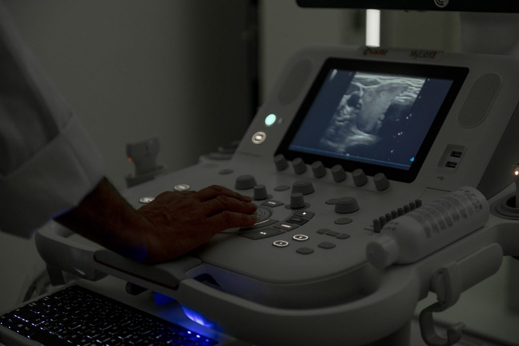
3D ULTRASOUND AND 4D
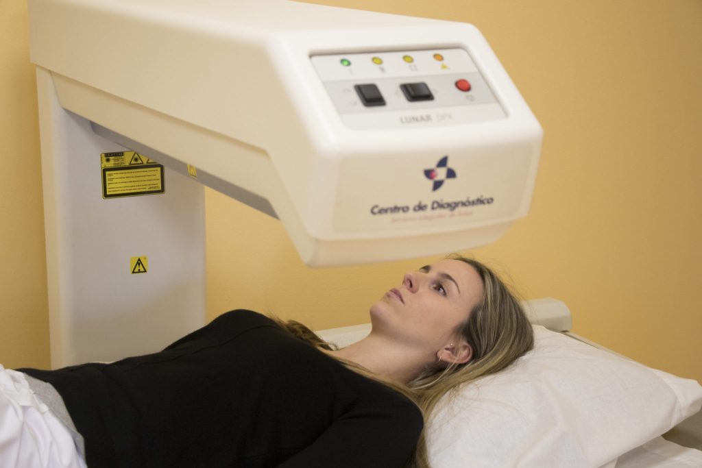
BONE DENSITOMETRY
BIOPSY STEREOTACTIC
MAGNETIC RESONANCE IMAGING OF THE BREAST
The magnetic resonance imaging of the breast it has been a big step forward in diagnostic imaging. Uses magnetic fields to generate internal signals of the body, captured by antennas special transform that information into images with high precision. In this technique, the breasts are kept in suspension and without compression, which improves the comfort and quality of the study.
Customarily used intravenous contrastbecause the malignant cells capture it with greater intensity, allowing you to identify areas suspicious. The images are processed in workstations advanced, which provide reconstructions three-dimensional and graphics capture detailed, which requires a thorough analysis by the specialist.
The major associations in oncology recommend this exploration in cases of carcinoma in situ, lobular hyperplasia, atypical, mammograms dense, or a personal history of breast cancer. It is also advisable for women with genetic mutations BRCA or inherited syndromes of high-risk (Li-Fraumeni, Cowden, Bannayan-Riley-Ruvalcaba), as well as in patients with high riskaccording to the assessment tools available medical.